


Vertigo, Dizziness
Third Window Syndrome: Symptoms, Diagnosis & Treatment
Third window syndrome: causes, symptoms like sound-induced dizziness and autophony, diagnosis and treatment. Book an appointment at EYiasis.
Secondary Endolymphatic Hydrops: Causes & Treatment
Secondary endolymphatic hydrops: causes, symptoms such as vertigo and tinnitus, diagnosis and treatment. Book an ENT neuro-otology visit at EYiasis.

Vestibular Migraine
Clinical experience has shown that vestibular migraine can be a chronic problem. Some patients report vertigo most days….

Balance. What controls the body’s position and posture?
Body balance is the ability to maintain your centre of gravity within your base of support, preventing falls and ensuring stability.

PPPD or Persistent Postural-Perceptual Dizziness
What is PPPD or Persistent Postural-Perceptual Dizziness? It is a condition which causes non-spinning dizziness and unsteadiness provoked by…

Vestibular Rehabilitation
Vestibular rehabilitation therapy (VRT) is a form of physical therapy that uses specialized exercises that result in gaze and gait stabilization…

What is Vestibular Rehabilitation?
Vestibular rehabilitation (VR) is a specialized form of therapy intended to alleviate both the primary and secondary problems caused by vestibular disorders…

Vertigo and Disequilibrium
Dizziness, vertigo and disequilibrium are common symptoms reported by many adults during visits to their doctors. A healthy vestibular system…

Vertigo, Dizzy spell, Balance Disorders
EYiasis Department of Vertigo and Balance Disorders addresses all patients, regardless of age, who suffer from dizziness, vertigo and unsteadiness.

Vertigo-Dizziness, what could be the problem!
Vertigo is a symptom, rather than a condition itself. It’s the sensation that you, or the environment around you, is moving or spinning.

Vertigo. Stop the room from spinning…
Vertigo, is a severe symptom that comes without warning and causes panic and disorientation. During episodes of vertigo the one unable to perform any activity

How to manage Vertigo and Dizziness
Vertigo and Dizziness: It is of utmost importance to be accurately diagnosed when symptoms of dizziness or vertigo are troubling you. Even though the…

Management of Vertigo-Dizziness
Treatment and management of vertigo depends on what’s causing it. Thus, after a thorough examination the otorhinolaryngologist will determine the etiology…

Vestibular Disorders and Vertigo
Vestibular disorders and Balance are determined by a complex combination of information flowing into the brain from the eyes, the labyrinths…
Introduction and Clarification of Clinical Terms: The Fundamental Difference Between Vertigo, Dizziness, and Unsteadiness
Vertigo, dizziness, and balance disorders are among the most common, concerning, and clinically complex symptoms that bring patients to a specialist otolaryngologist and neurotologist. In everyday communication, most people tend to use the words “vertigo” and “dizziness” interchangeably to describe any sensation of disorientation, unsteadiness, lightheadedness, or general physical malaise.
In medicine, however—and especially in neurotology—these terms describe entirely different clinical entities arising from different pathophysiological mechanisms and requiring fundamentally different diagnostic and therapeutic approaches. Accurate differentiation and a thorough understanding of these symptoms form the cornerstone of rational patient management, the avoidance of inappropriate medication, and definitive recovery.

Clear Clinical Differentiation of Symptoms
True Vertigo: Strictly defined as an illusion of movement, and more specifically an illusion of rotation. The patient experiences the intense and often frightening impression that either they or the environment around them is spinning, rotating, tilting, swaying, or turning upside down, even though their body remains completely still in bed or in a chair.
True rotational vertigo is almost always a pathognomonic sign of dysfunction affecting the vestibular system of the inner ear, the vestibular nerve, or the vestibular nuclei of the brainstem. In most cases, it is accompanied by reflex symptoms of autonomic nervous-system activation, including debilitating nausea, repeated vomiting, facial pallor, cold sweats, palpitations, and intense psychological panic, as well as objective findings such as nystagmus—the involuntary, rhythmic, rapid movement of the eyes.
Dizziness / Lightheadedness: Describes a non-spinning sensation of malaise, mental clouding (brain fog), “emptiness,” floating in space, feeling “drunk,” lightheadedness, or impending fainting (presyncope). Dizziness does not include the illusion that the surroundings are spinning and, as a rule, is not caused by disease of the inner-ear labyrinth.
More common causes include orthostatic hypotension, cardiovascular disorders, arrhythmias, hypoglycemia, severe anemia, adverse effects of antihypertensive or psychotropic medication, cervical syndrome, severe psychological stress, panic attacks, and chronic hyperventilation.
Disequilibrium & Balance Impairment: The patient describes a sense of insecurity, swaying, and loss of control over their center of gravity while standing or walking. They characteristically report feeling as though they are “stepping into empty space” or walking on “spongy ground”; they may stumble, adopt a wide-based gait, and tend to veer or fall to one side, without necessarily experiencing rotational vertigo or dizziness while seated or lying down.
This may be caused by chronic bilateral labyrinthine damage; neurological disorders of the central nervous system, such as cerebellar ataxia and Parkinson’s disease; impaired deep sensation (proprioception); or sensory peripheral neuropathy, as may occur with undiagnosed or poorly controlled diabetes mellitus.
Anatomy and Neurophysiology of the Balance System
Maintaining physical balance, spatial orientation, and visual-field stability during head movement results from the continuous, harmonious cooperation and integration of information from three distinct sensory systems, whose pathways converge and are processed within the central nervous system:
The Vestibular System of the Inner Ear (the Labyrinth): In addition to the cochlea, which is responsible for hearing, the inner ear contains the membranous and bony labyrinth of balance. The peripheral labyrinth comprises three semicircular canals—the horizontal, anterior, and posterior canals—which are filled with a specialized fluid called endolymph and are oriented at right angles to one another in the three dimensions of space.
The semicircular canals detect angular and rotational accelerations of the head with exceptional precision. The vestibular system also includes two otolith organs, the utricle and saccule, each containing a gelatinous membrane to which microscopic calcium-carbonate crystals—the otoliths or otoconia—are attached. The otolith organs detect gravity, head tilt, and rectilinear or linear acceleration, such as when traveling in an elevator, accelerating in a vehicle, or bending down.
- The Visual System: Vision provides the brain with visual references regarding the position and movement of the body in relation to the stable environment and the horizon. It is responsible for immediately detecting any displacement of the body in space.
- The Proprioceptive and Somatosensory System: Specialized mechanoreceptors and sensors located in the muscles, tendons, and joints—with particularly high density in the cervical spine, hips, knees, and soles of the feet—continuously inform the brain about the position of the limbs in space, joint angle, pressure exerted against the ground, and muscle tone.
All this sensory information is collected and processed instantaneously by the vestibular nuclei of the brainstem and the cerebellum, which function as the body’s central computer. Provided that the information from the labyrinth, eyes, and joints agrees completely, a person maintains perfect balance without any conscious effort.
When damage, inflammation, or dysfunction occurs in one of these systems—for example, when the right labyrinth becomes acutely inflamed and stops sending signals while the left labyrinth continues to function normally—the brain receives conflicting, asymmetric information. The eyes and legs indicate that the body is motionless, but the asymmetry of the labyrinthine signals is interpreted by the brain as violent rotation of the body.
This sensory conflict immediately triggers the clinical picture of vertigo, produces reflex eye movements (nystagmus), and stimulates the vomiting centers in the medulla oblongata.
Pathophysiology, Causes, and Predisposing Factors of Vertigo and Dizziness
Etiological classification of vertigo is fundamental in neurotology and is divided into two broad categories: peripheral lesions, which affect only the inner ear and vestibular nerve and account for the majority of vertigo cases; and central lesions, which affect the central nervous system, brainstem, or cerebellum and require prompt, meticulous diagnostic evaluation.
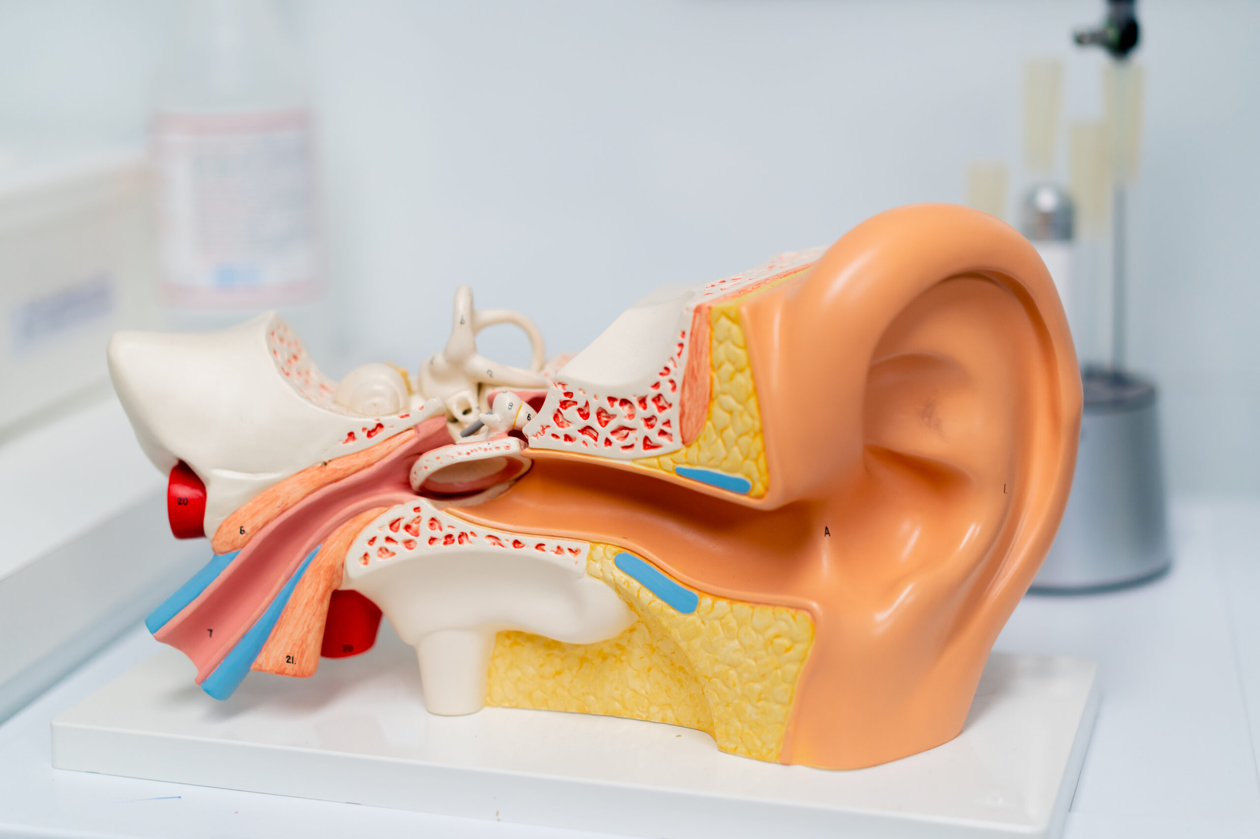
Peripheral Causes of Vertigo (Diseases of the Inner Ear and Labyrinth)
Peripheral vertigo usually produces a more intense and dramatic clinical presentation and has a clear relationship to head movement, but it rarely threatens the patient’s life and carries an excellent prognosis and high cure rates with appropriate treatment.
Benign Paroxysmal Positional Vertigo (BPPV): This is the most common cause of vertigo worldwide and accounts for a very large proportion of patients presenting with a vertigo attack. Its pathophysiology (known as canalithiasis or cupulolithiasis) involves microscopic calcium-carbonate crystals—the otoliths—becoming dislodged from their normal position in the utricle.
These free-floating crystals enter the endolymph within one of the semicircular canals—in the overwhelming majority of cases, the posterior canal, and less often the horizontal or anterior canal. When the patient changes position, such as when lying down, turning over in bed, bending forward, or extending the head backward to look up, gravity causes the crystals to move within the canal.
This movement creates an abnormal flow of endolymph that deflects the sensory organ (the cupula) and sends a false signal of violent rotation to the brain. The clinical picture is characterized by sudden, brief attacks of rotational vertigo lasting less than one minute and accompanied by nausea, without ever causing hearing loss, tinnitus, or neurological deficits.
Ménière’s Disease (Endolymphatic Hydrops): A chronic, recurrent, idiopathic disorder of the inner ear caused by an abnormal increase in the volume and pressure of endolymph within the membranous labyrinth and cochlea. This excessive fluid pressure (endolymphatic hydrops) distends the internal membranes, which periodically develop microscopic ruptures.
This leads to mixing of endolymph and perilymph, toxic disruption of the sensory hair cells, and transient interruption of auditory and vestibular function. Ménière’s disease presents with a characteristic clinical tetrad of symptoms in the affected ear:
- Spontaneous, recurrent attacks of severe rotational vertigo lasting from twenty minutes to several hours and accompanied by intense nausea, vomiting, and pallor.
- Fluctuating sensorineural hearing loss, which affects the low and middle frequencies in the early stages of the disease and often improves after the attack ends, but becomes permanent and worsens over the years.
- Tinnitus in the affected ear, usually low-frequency (a buzzing sound resembling wind, machinery, or whistling), which intensifies dramatically before and during the vertigo attack.
- A sensation of fullness, pressure, heaviness, or “blockage” deep inside the ear.
Vestibular Neuritis / Vestibular Neuronitis: An acute, benign but exceptionally severe and dramatic inner-ear disorder characterized by sudden, complete or partial loss of function of the peripheral vestibular nerve on one side. It is almost always caused by a viral infection or reactivation of a latent neurotropic virus, such as herpes simplex virus, influenza viruses, varicella-zoster virus, or adenoviruses, within Scarpa’s vestibular ganglion.
Acute edema and ischemic inflammation of the nerve abruptly interrupt the balance signals traveling from the affected ear to the brain. The patient experiences sudden, continuous, unremitting, and extremely severe rotational vertigo lasting uninterruptedly for days, accompanied by uncontrollable vomiting, marked spontaneous nystagmus, inability to stand, and a tendency to fall toward the affected side.
The fundamental diagnostic feature distinguishing vestibular neuritis from other disorders is the complete absence of audiological symptoms: the patient’s hearing remains 100% intact, with no hearing loss or tinnitus.
Labyrinthitis: This is simultaneous acute inflammation of the entire inner ear, involving both its vestibular and cochlear components. It may be caused by a viral infection or a bacterial infection, often occurring as a complication of neglected acute otitis media, chronic otitis media with cholesteatoma, or meningitis.
Its clinical presentation is identical to vestibular neuritis in terms of prolonged, violent vertigo, vomiting, and unsteadiness, but is clearly distinguished by concurrent severe sensorineural hearing loss, intense tinnitus, and often ear pain in the affected ear.
Perilymphatic Fistula: An abnormal tear or breach in the delicate membranes separating the middle and inner ear (the round or oval window), resulting in leakage of perilymph from the inner-ear cavity into the middle-ear cavity. It is usually caused by significant barotrauma, such as deep diving or a rapid aircraft descent; direct head trauma; intense physical exertion that raises intracranial pressure, such as lifting excessive weight, forceful coughing, or sneezing; or a complication of ear surgery, such as stapedectomy for otosclerosis.
It causes episodes of vertigo and unsteadiness characteristically triggered by increased pressure (Hennebert’s sign) or exposure to loud sounds (Tullio phenomenon), accompanied by fluctuating hearing loss.
Acoustic Neuroma (Vestibular Schwannoma): A slow-growing, benign tumor arising from the Schwann cells forming the sheath of the vestibular branch of the eighth cranial nerve, within the internal auditory canal or at the cerebellopontine angle. Because the tumor grows extremely slowly, the brain has time to activate central compensatory mechanisms in response to the reduced vestibular signal.
For this reason, an acoustic neuroma rarely presents with acute, dramatic rotational vertigo. Its classic symptoms are progressive, unilateral, asymmetric sensorineural hearing loss—particularly at high frequencies—persistent unilateral tinnitus, and a chronic, vague sense of unsteadiness, dizziness, and uncertainty while walking. At an advanced size, it may compress adjacent cranial nerves and cause facial numbness through involvement of the trigeminal nerve or facial weakness through involvement of the facial nerve.
- Other Peripheral Causes: Otosclerosis involving the cochlea, congenital anomalies of the inner ear, and superior semicircular canal dehiscence syndrome (SSCD), which causes sound-induced vertigo, autophony, and bone-conduction hyperacusis.
Central Causes of Vertigo (Diseases of the Central Nervous System)
Vertigo of central origin may produce a less dramatic spinning sensation, but it is accompanied by prominent gait unsteadiness, follows a continuous and unremitting course, is not readily suppressed by visual fixation, and may conceal life-threatening conditions requiring immediate medical intervention.
Vestibular Migraine / Migrainous Vertigo: This is the second most common cause of recurrent vertigo worldwide (after BPPV) and the most common central cause of vestibular dysfunction. Its pathophysiology is associated with abnormal neuronal hyperexcitability, vasoconstriction, and neurogenic vascular inflammation in the brainstem, cerebellum, and pathways supplying the labyrinth.
It presents with episodes of vertigo, severe dizziness, or unsteadiness lasting from five minutes to as long as three full days. Its crucial clinical feature is that the vertigo does not always need to be accompanied by an active, severe headache—it often presents as migraine without headache (silent migraine).
The patient nevertheless has a history of migraine, and episodes of dizziness are accompanied by classic migrainous symptoms such as hypersensitivity to light (photophobia) and noise (phonophobia), visual aura with flashes or zigzag patterns, and marked intolerance and dizziness in moving, visually complex environments (visual vertigo).
Stroke (Ischemia or Hemorrhage of the Brainstem and Cerebellum): Acute occlusion or hemorrhage affecting vessels of the posterior cerebral circulation, such as the anterior inferior cerebellar artery (AICA), posterior inferior cerebellar artery (PICA), or basilar artery, causes ischemic infarction in the vestibular nuclei or cerebellum.
This is a life-threatening emergency requiring immediate transfer to hospital. It differs from uncomplicated peripheral vertigo because it is accompanied by central neurological deficits such as diplopia (double vision), dysarthria, dysphagia, severe cerebellar ataxia with inability to stand, and altered sensation, numbness, or weakness in the face and limbs.
- Vertebrobasilar Insufficiency (VBI): A transient reduction in blood flow through the vertebral and basilar arteries due to atherosclerosis, vasospasm, or mechanical compression by cervical osteophytes. It causes brief, recurrent episodes of vertigo and lightheadedness, often triggered by hyperextension or sudden rotation of the neck, and may be accompanied by transient blurred vision, falling to the ground without loss of consciousness (drop attacks), or weakness in the legs.
- Demyelinating Diseases (Multiple Sclerosis): The development of demyelinating plaques along the neural connections of the vestibular system within the brainstem and cerebellum is a common cause of vertigo and unsteadiness in young adults and may be the first clinical manifestation of disease onset.
- Other Central Disorders: Tumors of the brainstem and fourth ventricle, neurodegenerative diseases of the central nervous system, Parkinson’s disease, Arnold–Chiari syndrome, and post-concussion syndrome following concussion or traumatic brain injury.
Systemic, Metabolic, and Functional Causes of Dizziness and Unsteadiness
In many cases, dizziness originates neither in the inner-ear labyrinth nor from organic brain damage, but from broader systemic, vascular, musculoskeletal, or psychosomatic disorders:
Cervicogenic Vertigo and Cervicogenic Dizziness: Caused by pathology of the cervical spine, such as cervical syndrome, degenerative spondyloarthropathy, herniated intervertebral discs, whiplash injuries following road-traffic accidents, or severe, chronic muscle spasm and shortening of the paraspinal, trapezius, and suboccipital muscles. The cervical region is exceptionally rich in proprioceptive mechanoreceptors, which convey information about head position to the brain.
When the cervical muscles are inflamed or in spasm, they send abnormal, distorted proprioceptive signals to the vestibular nuclei that conflict with the correct signals from the ears and eyes. The patient experiences continuous, vague dizziness, clouding, a “floating” sensation, and heaviness at the back of the head and neck, which worsen with neck movements, anxiety, and prolonged poor posture in front of screens.
Orthostatic Hypotension and Cardiovascular Disorders: A sudden, significant fall in blood pressure when the patient rises from a seated or lying position to stand causes a transient reduction in cerebral perfusion. The patient immediately experiences dimming of vision (“everything goes black”), a sense of emptiness, tinnitus, dizziness, weakness in the lower limbs, and a feeling of impending fainting, which resolves as soon as they lie down.
Similar dizziness may be caused by cardiac arrhythmias such as atrial fibrillation and tachycardia, as well as by heart failure or valvular heart disease.
Drug Toxicity and Iatrogenic Dizziness: Dizziness is one of the most common adverse effects of medication, particularly in older patients taking multiple drugs. Antihypertensive medicines, diuretics, and vasodilators cause orthostatic dizziness. Specific classes of medicines known as ototoxic drugs—including aminoglycoside antibiotics (gentamicin, amikacin), loop diuretics (furosemide), and certain chemotherapeutic agents (cisplatin)—directly destroy the sensory hair cells of the labyrinth, causing permanent bilateral vestibular damage, severe unsteadiness, oscillopsia, and significant hearing loss.
Psychotropic medicines, anxiolytics, benzodiazepines, hypnotics, antidepressants, and antiepileptic drugs also reduce neural transmission and cause a feeling of intoxication, fatigue, drowsiness, and unsteadiness.
Persistent Postural-Perceptual Dizziness (PPPD): A contemporary functional neurotological disorder formally recognized by the Bárány Society and the most common cause of chronic, daily dizziness lasting more than three months. It is usually initially triggered by a genuine acute episode of vertigo (such as vestibular neuritis, BPPV, or a Ménière’s attack), or by a panic attack, acute stress, or concussion.
Even after the initial organic inner-ear lesion has healed completely, the patient’s brain “fails” to return to normal, automatic, subconscious control of balance. The balance system remains trapped in a state of hypervigilance and hypersensitivity; the patient becomes excessively dependent on visual information and anxiously monitors every slight sway of the body.
The patient experiences an unremitting, daily sensation of swaying, vague dizziness, and “floating” in space, which worsens dramatically while standing, during physical movement, and in environments with intense visual motion and crowds, such as supermarkets, shopping centers, busy streets, and digital screens.
Clinical Features: Presentation and Red Flags
The clinical presentation of vestibular disorders is not confined to spinning and rotational sensations; it spans a broad, complex spectrum of physical, sensory, audiological, autonomic, and psychological symptoms that can completely disrupt the patient’s everyday life:

- Sensory and Visual Symptoms: The patient experiences rotational vertigo or sensations of hovering, sinking, floating, overturning, or swaying. They often report oscillopsia—the distressing visual impression that stationary objects around them bounce, shake, or move rhythmically with every step or head movement because of loss of the vestibulo-ocular reflex (VOR).
- Autonomic Nervous-System Symptoms: The close anatomical and neuronal proximity of the vestibular nuclei to the vomiting and autonomic centers in the medulla oblongata causes debilitating nausea, repeated vomiting, pallor, cold sweating, excessive salivation, tachycardia, and a profound sense of physical exhaustion.
- Motor Symptoms and Unsteadiness: Manifestations include unsteadiness when walking, stumbling, inability to maintain a straight course, widening the stance for greater support, a tendency to veer or fall to one side—usually toward the affected labyrinth—and an inability to stand in darkness or on uneven ground.
- Audiological Symptoms: In disorders affecting the entire inner ear, vertigo is accompanied by unilateral or bilateral hearing loss, severe tinnitus (buzzing, whistling, machinery, wind, or ringing sounds), a sensation of fullness, pressure, and blockage deep in the ear, and intolerance to loud sounds (hyperacusis).
- Psychological and Cognitive Effects: Patients troubled by recurrent or chronic vertigo develop severe anxiety, panic attacks, agoraphobia, and avoidance of going out because of the persistent fear of another unpredictable attack or a fall in public. They also report mental clouding (brain fog), difficulty concentrating, easy fatigability, and an inability to focus at work or while reading.
Critical Warning Signs: When Vertigo Is a Medical Emergency (Red Flags)
Although in the overwhelming majority of cases vertigo is caused by benign peripheral ear disorders that are not life-threatening, specific clinical warning signs—the so-called red flags—indicate involvement of the central nervous system and a possible stroke or brainstem hemorrhage. The presence of any of the following symptoms requires the patient’s immediate emergency transfer to hospital for neurological assessment and imaging:
- Sudden, first-ever, extremely severe vertigo in a patient with cardiovascular risk factors such as arterial hypertension, diabetes mellitus, smoking, dyslipidemia, advanced age, or a history of arrhythmias.
- A sudden, thunderclap headache of unprecedented intensity accompanying the vertigo (“the worst headache of my life”).
- The onset of diplopia (double vision), sudden blurred vision, or visual-field loss.
- Dysarthria, dysphonia, “slurred” speech, inability to articulate speech, or difficulty finding words.
- Dysphagia, difficulty swallowing liquids or solid food, and a sensation of choking.
- Severe, prominent cerebellar ataxia in which the patient cannot stand by any means or take even a single step without collapsing to the ground (“falls like a log”).
- Altered sensation, numbness, reduced sensation, weakness, paresis, or paralysis affecting half of the face, an arm, a leg, or an entire side of the body.
- Loss of consciousness, a fainting episode, generalized confusion, lethargy, or a seizure.
- Sudden, simultaneous, complete deafness in both ears.
Diagnostic Approach and Specialist Neurotological Assessment at “EViasis”
Modern neurotological diagnosis of vertigo and dizziness has definitively abandoned the vague and dangerous practice of blindly prescribing “dizziness pills” without an accurate diagnosis. At the specialist Neurotology Department of the “EViasis” Model Medical Center in Marousi, diagnosis is based on a rigorous, comprehensive, scientifically documented clinical and laboratory protocol using state-of-the-art diagnostic systems:
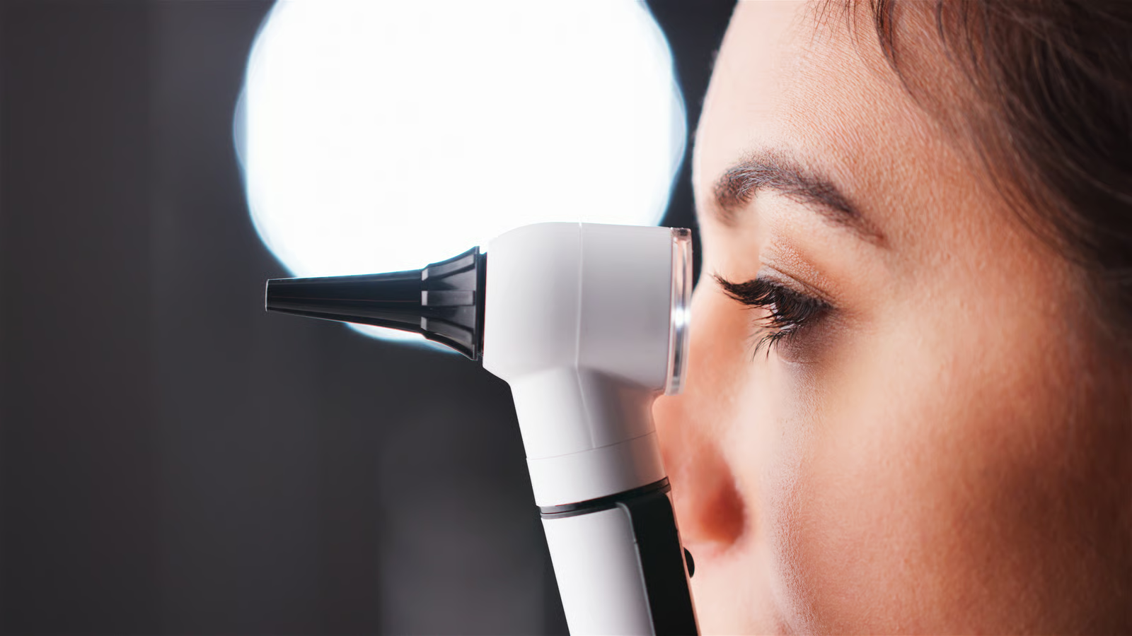
Detailed, Targeted Neurotological History
The patient’s history is the most important diagnostic tool, allowing an experienced neurotologist to establish the correct diagnosis in the majority of cases. The physician meticulously investigates the precise nature of the symptom—whether it is rotation, clouding, or unsteadiness; the exact duration of each attack—whether seconds as in BPPV, minutes to hours as in Ménière’s disease, or continuous days as in vestibular neuritis; the factors that trigger symptoms, such as positional changes, coughing, standing, loud sounds, or stress; the presence of audiological symptoms; and the patient’s medical history, including migraine, viral infections, cervical symptoms, cardiovascular disease, and current medication.
Bedside Clinical Neurotological Examination
The patient undergoes a meticulous ENT, audiological, and clinical neurotological examination:
- Otomicroscopy and Endoscopy: Detailed examination of the tympanic membrane and external auditory canal under magnification to exclude local inflammation, perforation, cholesteatoma, middle-ear fluid, or obstruction by impacted cerumen.
- Comprehensive Audiological Assessment: Pure-tone and speech audiometry in a dedicated sound-treated booth, tympanometry, and acoustic-reflex testing to assess cochlear function and detect lesions affecting the auditory nerve, as in Ménière’s disease and acoustic neuroma.
- The Clinical HINTS Protocol (Head-Impulse, Nystagmus, Test-of-Skew): An exceptionally powerful, revolutionary diagnostic protocol comprising three clinical tests in the hands of a specialist neurotologist. In patients presenting with an acute, continuous, unremitting vertigo syndrome, the HINTS protocol has greater diagnostic sensitivity and accuracy (100%) than brain magnetic resonance imaging during the first 24 to 48 hours for differentiating a peripheral lesion (vestibular neuritis) from a central vascular lesion (brainstem or cerebellar infarction):
- Head-Impulse Test (HIT): The physician asks the patient to fix their gaze on the physician’s nose and performs rapid, unpredictable, small rotational movements of the patient’s head to the right and left. If a corrective saccadic eye movement appears, the examination confirms a peripheral lesion in the corresponding labyrinth (vestibular neuritis). If the test is completely normal in a patient with continuous, violent vertigo, this is a critical warning sign of cerebral infarction.
- Assessment of Nystagmus: Spontaneous and provoked nystagmus is assessed in all gaze positions. Nystagmus of peripheral origin is horizontal-torsional, always beats in one fixed direction, and is suppressed by visual fixation on a stationary point. By contrast, purely vertical nystagmus (upbeat or downbeat), nystagmus that changes direction according to gaze position, and nystagmus that is not suppressed by vision indicate a central lesion of the central nervous system.
- Alternate Cover Test (Test of Skew): Vertical ocular alignment is assessed. Detection of a vertical strabismic deviation, in which one eye moves vertically when uncovered, is a clear indication of a brainstem lesion.
- Specialist Positional Tests:
- Dix–Hallpike Test and Side-Lying Test: These are the gold standard for diagnosing BPPV of the posterior semicircular canal. The physician rapidly moves the patient from a seated to a supine position, with the head turned 45 degrees and hanging backward, while observing the latency, onset, and fatigability of the characteristic transient torsional-upbeating nystagmus.
- Supine Roll Test (Pagnini–McClure Test): Performed with the patient lying down while the head is turned 90 degrees to the right and left to diagnose BPPV of the horizontal semicircular canal.
- Static and Dynamic Balance and Gait Tests:
- Romberg Test and Foam Romberg Test: Body sway is assessed while the patient stands with feet together, first with eyes open and then closed, both on a firm surface and on a specialized foam pad that removes proprioceptive input from the soles of the feet.
- Unterberger–Fukuda Test: The patient marches in place for fifty steps with eyes closed and arms extended forward. Involuntary rotation of the body by more than thirty degrees indicates a peripheral lesion and reduced labyrinthine function on the side toward which the patient turns.
- Tandem (Heel-to-Toe) Gait Test: The patient walks in a straight line, placing the heel of one foot directly in front of the toes of the other, to assess cerebellar coordination.
- Assessment of Cranial Nerves and Cerebellar Function: Eye movements and the trigeminal and facial nerves are assessed, together with finger-to-nose and heel-to-shin testing and evaluation of rapid alternating movements.
Laboratory Neurotological, Audiological, and Imaging Assessment
When the clinical examination indicates the need for more extensive investigation, “EViasis” offers and performs advanced diagnostic tests to map the vestibular system comprehensively:
- Videonystagmography (VNG) and Electronystagmography (ENG): An objective digital recording of eye movements and nystagmus using specialized goggles fitted with infrared cameras. The examination includes oculomotor and positional tests and the gold-standard caloric test, in which each labyrinth is stimulated separately in a controlled manner by introducing warm and cool air or water into the external auditory canal. Caloric testing measures the percentage of hypofunction, reduced function, or paresis in each labyrinth with mathematical precision.
- Video Head Impulse Test (v-HIT): A state-of-the-art, painless, high-speed examination that assesses the function of all six semicircular canals of the inner ears individually and with absolute precision in a matter of minutes. It can detect extremely subtle, hidden saccadic eye movements (overt and covert saccades) that are not visible to the naked eye.
- Vestibular Evoked Myogenic Potentials (VEMPs): An electrophysiological examination—cervical c-VEMPs and ocular o-VEMPs—that assesses the integrity and function of the inner ear’s otolith organs (utricle and saccule) and of the superior and inferior vestibular nerves. It is valuable in diagnosing Ménière’s disease and superior canal dehiscence syndrome (SSCD).
- Computerized Dynamic Posturography / Dynamic Balance Assessment: Objectively evaluates the contribution and adequacy of the vestibular, visual, and proprioceptive systems in maintaining physical balance under different sensory conditions.
Imaging (Magnetic Resonance Imaging and Computed Tomography): When there are indications of central pathology, asymmetric hearing, tinnitus, or failure to respond to treatment, high-resolution magnetic resonance imaging of the brain and petrous temporal bones (MRI/MRA) is performed using a dedicated thin-slice inner-ear protocol and a paramagnetic contrast agent (gadolinium).
The aim is to exclude acoustic neuroma, vascular lesions of the brainstem, multiple sclerosis, or tumors. Carotid and vertebral artery duplex ultrasonography may also be requested to assess blood flow, along with specialized hematological and metabolic testing (vitamin D, vitamin B12, glucose, and thyroid hormones).
Treatment: From the Acute Phase to Definitive Recovery and Rehabilitation
Treatment of vertigo and dizziness at “EViasis” is never one-dimensional or empirical; it is strictly etiological, individualized, and tailored to each patient’s specific needs, combining every contemporary medical option:

Therapeutic Repositioning Maneuvers for BPPV
Benign paroxysmal positional vertigo is never treated with medication, and anti-vertigo drugs are entirely ineffective and harmful in this condition. The treatment of choice—with success rates reaching 90–95% after the very first session—is the immediate performance of specialized, painless mechanical repositioning maneuvers by the specialist physician on the examination couch:
- Epley Maneuver and Semont Maneuver: Used to reposition particles in BPPV of the posterior semicircular canal. The physician guides the patient’s head and body through a specific sequence of positions based on gravity, allowing the dislodged crystals to travel through the entire semicircular canal and return to their normal position in the utricle.
- Barbecue Roll (Lempert Maneuver) and Gufoni Maneuver: Performed to reposition the crystals when the horizontal semicircular canal is affected.
- Yacovino Maneuver: Used for the rare involvement of the anterior semicircular canal.
- After the maneuver has been performed successfully, the vertigo resolves immediately, the crystals return to their proper position, and the physician provides instructions to avoid sudden head movements over the next few days so that they remain stabilized.
Pharmacological Treatment (Acute Phase, Regulation, and Maintenance)
Pharmacotherapy has clear, specific, and time-limited medical indications:
During an Acute Attack (First 1–3 days of acute, violent vertigo, e.g. vestibular neuritis or a Ménière’s attack): Vestibular suppressants are administered to relieve severe nausea, uncontrollable vomiting, and the violent spinning sensation. Antihistamines (dimenhydrinate/Dramamine, meclizine), antiemetic and neuroleptic drugs (metoclopramide, ondansetron, sulpiride/Dogmatyl, domperidone), and short courses of benzodiazepines (diazepam, lorazepam, clonazepam) are used.
In addition, in cases of acute vestibular neuritis, labyrinthitis, or sudden hearing loss, a corticosteroid regimen is administered promptly (oral cortisone or intratympanic dexamethasone injections) to reduce nerve edema and protect hearing.
- Critical Medical Warning: Vestibular suppressants (such as dimenhydrinate and benzodiazepines) must be discontinued after the first 48 to 72 hours. Prolonged, chronic use suppresses the brain’s natural neuroplasticity, prevents the development of central compensation, and turns a transient labyrinthine problem into permanent, debilitating chronic unsteadiness and dependence.
- For Long-Term Regulation, Maintenance, and Relapse Prevention:
- Betahistine (Serc, Histimed): A potent, specific vasodilatory and neuromodulatory agent for the inner ear that improves labyrinthine microcirculation, regulates endolymphatic pressure in Ménière’s disease, and significantly reduces the frequency and severity of attacks.
- Diuretics: Acetazolamide or a hydrochlorothiazide/amiloride combination to reduce fluid volume and relieve endolymphatic hydrops in Ménière’s disease.
- Preventive Treatment for Vestibular Migraine: Beta blockers (propranolol), calcium-channel blockers (flunarizine), anticonvulsant/antiepileptic drugs (topiramate, valproic acid), specific antidepressants (SSRIs/SNRIs), and targeted supplements of magnesium, riboflavin (vitamin B2), and coenzyme Q10 are administered.
Vestibular Rehabilitation Therapy (VRT) Program
Vestibular Rehabilitation Therapy (VRT) is a highly specialized, non-invasive, scientifically documented therapeutic method using specific physical and oculomotor exercises. It is the cornerstone of treatment for patients with chronic persistent unsteadiness, recovering vestibular neuritis, functional dizziness (PPPD), Ménière’s disease between attacks, and older people at high risk of falls.
The method is based on the principles of brain neuroplasticity and central compensation. Through a personalized program, the brain is retrained to process balance signals correctly, disregard deficient information from the affected labyrinth, and make greater use of visual and proprioceptive references:
- Gaze Stabilization Exercises (Cawthorne–Cooksey Exercises and X1/X2 Viewing): The patient fixes their gaze on a stationary visual target while rhythmically moving the head along the horizontal and vertical axes. This retrains the vestibulo-ocular reflex (VOR), restores visual-field stability, and eliminates oscillopsia.
- Habituation and Desensitization Exercises: The patient is repeatedly, gradually, and in a controlled manner exposed to the exact head movements and visual stimuli that provoke dizziness (such as bending down, sudden turns, or the visual motion in a supermarket), causing the brain to become desensitized and reduce its panic response.
- Static and Dynamic Balance, Proprioception, and Gait Exercises: Training to maintain balance on stable and unstable surfaces and foam mats with eyes open and closed; dynamic walking practice combined with head movements, turns, and obstacles; and exercises to strengthen the muscles of the lower limbs and trunk.
Medical Acupuncture and Auricular Acupuncture for Vertigo, Dizziness, and Tinnitus at “EViasis”
Medical acupuncture and auricular acupuncture, performed by the Scientific Director of “EViasis” Dr Christina Efthymiou, integrate contemporary Western neurotology with the holistic medical power of neuromodulation. They constitute an excellent, entirely safe complementary treatment with high clinical effectiveness for patients with vertigo, chronic unsteadiness, cervicogenic vertigo, vestibular migraine, tinnitus, and functional dizziness (PPPD):
- Enhancement of Microcirculation and Oxygenation: The painless placement of specialized, sterile, single-use needles at targeted anatomical and neurovascular points causes local vasodilation of small blood vessels, rapidly increasing blood flow and oxygen delivery and promoting the clearance of toxic metabolites in the inner-ear labyrinth, cochlea, vestibular nerve, and brainstem. This effect is fundamental to controlling attacks in Ménière’s disease and relieving tinnitus.
- Release of Cervical Muscle Spasm and Cervicogenic Vertigo: Body acupuncture and electroacupuncture effectively decompress and release chronic muscle spasms and myofascial trigger points in the suboccipital, paraspinal, sternocleidomastoid, and trapezius muscles of the cervical region. In this way, they definitively stop the abnormal, distorted proprioceptive signals responsible for cervicogenic dizziness and unsteadiness.
Regulation of the Autonomic Nervous System, Anxiety, and PPPD: Stimulation of the parasympathetic nervous system—and specifically branches of the vagus nerve, through both body acupuncture and auricular acupuncture on the pinna—immediately triggers the release of endorphins and serotonin, alleviates nausea, stabilizes the heart rate, drastically reduces internal stress, and eliminates the central nervous system’s state of hypervigilance.
This effect is invaluable in freeing patients from the vicious cycle of chronic anxiety and persistent functional dizziness (PPPD).
Surgical and Interventional Options (for Refractory Cases)
In an exceptionally small proportion of patients (fewer than 5% overall) who have severe Ménière’s disease refractory to conservative treatment with debilitating vertigo attacks, recurrent and unbearable BPPV, or a large acoustic neuroma, interventional and surgical options are used:
- Intratympanic Gentamicin Injections (Chemical Labyrinthectomy): A controlled dose of an ototoxic antibiotic is injected into the middle ear to selectively and partially destroy vestibular cells in the affected ear, permanently stopping vertigo attacks in Ménière’s disease.
- Endolymphatic Sac Decompression: A hearing-preserving operation intended to drain excess fluid and reduce pressure in the inner ear.
- Vestibular Neurectomy: Surgical division of the vestibular branch of the eighth cranial nerve within the skull, eliminating vertigo while preserving 100% of the patient’s existing hearing.
- Labyrinthectomy: Surgical removal of the labyrinth, performed exclusively in patients who have already completely lost hearing in the affected ear.
- Surgical Excision or Radiosurgery (Gamma Knife) for Acoustic Neuroma.
Prevention, Dietary Guidance, and Quality-of-Life Advice
Appropriate daily prevention, targeted dietary adjustments, and healthy habits play a decisive role in maintaining labyrinthine homeostasis, preventing relapses, and meaningfully improving quality of life:

Dietary Guidance for Ménière’s Disease and Endolymphatic Hydrops
- Strict Salt (Sodium) Restriction: Sodium causes marked fluid retention in the body and directly increases the pressure and volume of endolymph within the inner ear, making it the principal trigger for vertigo attacks. Daily sodium intake should not exceed 1,500–2,000 mg. Table salt, cheeses, processed meats, salt-cured foods, olives, pickles, canned foods, salty snacks, ready-made sauces, stock cubes, and processed foods should be avoided.
- Consistent, Adequate Hydration: Drink two to two and a half liters of plain water daily, distributed evenly throughout the day, to avoid major fluctuations in the inner ear’s fluid balance.
- Limit Caffeine, Alcohol, Chocolate, and Sugar: Caffeine (coffee, energy drinks, and black tea) and alcohol cause vasoconstriction and disrupt microcirculation and nerve conduction in the labyrinth, worsening vertigo and tinnitus.
- Smoking Cessation: Nicotine causes severe, prolonged spasm of the capillaries supplying the labyrinth and auditory nerve.
Ergonomics, Neck Care, and Stress Management
- Avoid prolonged poor positioning of the head and cervical spine in front of computer and mobile-phone screens (text-neck syndrome). Adjust the computer screen so that it is exactly at eye level.
- Use a suitable anatomical sleeping pillow that supports the neck in a neutral position, preventing hyperextension or excessive flexion during sleep.
- Perform gentle daily stretches of the cervical and trapezius muscles during work and avoid sudden, forceful head rotations.
- Manage psychological stress through relaxation techniques, appropriate breathing, and a consistent, adequate night-time sleep schedule of seven to eight hours.
Fall Prevention and Safety at Home (for Older People and Patients with Unsteadiness)
- Remove loose rugs, cables, and small pieces of furniture from hallways in the home to prevent trips.
- Install adequate automatic night lighting in hallways and the bathroom. A patient with a vestibular disorder relies completely on vision for support and is at serious risk of falling when walking in the dark.
- Install non-slip mats and support grab rails in the bath and shower.
- Include the patient in a program of regular, low-intensity aerobic exercise, such as walking, swimming, or Tai Chi, which strengthens muscles, enhances proprioception, and accelerates central cerebral compensation.
About the Doctor and the Specialist “EViasis” Medical Center
The “EViasis” Model Medical Center for Otorhinolaryngology, Neurotology & Medical Acupuncture (ent.gr), based in Marousi, Attica, is a contemporary center of excellence for the thorough diagnosis and comprehensive treatment of vertigo, dizziness, tinnitus, and all balance disorders.
The Scientific Director of “EViasis,” Dr Christina Efthymiou, is an ENT surgeon, specialist neurotologist, and certified medical acupuncturist with extensive clinical, hospital, surgical, and research experience at leading medical centers in Greece and abroad. She has received advanced training and specialization in inner-ear disorders, neurotology, vestibular rehabilitation, medical acupuncture, and auricular acupuncture, harmoniously combining conventional Western medicine with contemporary holistic neuromodulatory treatments.

The philosophy of “EViasis” places the patient and their individual needs at its absolute center. Dr Christina Efthymiou approaches patients with vertigo, dizziness, and unsteadiness without superficial assessments or hasty, indiscriminate prescribing. She devotes all the clinical time required to take a detailed history and conduct specialist neurotological, digital, and audiological testing, seeking to identify the true root of the problem.
Why Choose “EViasis” for Vertigo and Dizziness in Marousi
- Complete Specialization in Neurotology: Thorough investigation, differential diagnosis, and management of the entire spectrum of peripheral and central vestibular disorders.
- State-of-the-Art Diagnostic and Laboratory Equipment: Specialist diagnostic testing can be performed promptly at one location, including videonystagmography (VNG), video head impulse testing (v-HIT), comprehensive audiological assessment, and tympanometry.
- Immediate Performance of Therapeutic BPPV Repositioning Maneuvers: A definitive, painless solution for benign paroxysmal positional vertigo within minutes in the examination room, without unnecessary and harmful medication.
- Individualized Vestibular Rehabilitation and Acupuncture Protocols: A unique combined therapeutic approach for refractory, chronic patients with persistent dizziness (PPPD), Ménière’s disease, cervicogenic vertigo, tinnitus, and stress.
- Comprehensive, Continuous Medical Support: Monitoring throughout the patient’s treatment journey until physical stability, well-being, and self-confidence are fully and definitively restored.
Access, Contact, and Patient Services
The “EViasis” Model Medical Center is located in a modern, comfortable, and easily accessible area of Marousi, Attica, serving patients daily both from Marousi and from across the Northern Suburbs, including Kifisia, Chalandri, Vrilissia, Melissia, Pefki, Lykovrysi, Psychiko, Filothei, Neo Irakleio, and Agia Paraskevi, as well as from throughout the Athens metropolitan area. For detailed information about the available Neurotology, Otorhinolaryngology, and Medical Acupuncture services, and to schedule your personal appointment with Dr Christina Efthymiou, please visit the practice’s official website at https://ent.gr/.
Comprehensive Frequently Asked Questions (FAQ)
The following is a detailed, evidence-informed medical guide answering more than fifteen common patient questions and addressing every likely concern about vertigo, dizziness, balance disorders, and their clinical management at “EViasis.”

What is the essential difference between true vertigo and ordinary dizziness?
Dizziness/lightheadedness is a general, vague sensation of malaise, mental clouding, feeling “drunk,” “floating” in space, unsteady, or about to faint, but without the patient feeling that the surroundings are spinning. It may be caused by low blood pressure, cervical syndrome, anxiety, anemia, hypoglycemia, or adverse effects of medication.
True vertigo, by contrast, is strictly the illusion of rotation and spinning. The patient sees and feels either themselves or the room around them spinning violently, as though on a spinning wheel or amusement-park ride, even though they remain completely still. True vertigo is almost always an indication of pathology affecting the vestibular system of the inner ear (the labyrinth) or the brain’s vestibular centers.
What is the most common cause of vertigo, and how is it treated?
The most common cause of vertigo worldwide, accounting for a very large proportion of all cases, is benign paroxysmal positional vertigo (BPPV). It is caused by microscopic crystals (otoliths) becoming dislodged into the endolymph within the semicircular canals of the inner ear.
It presents with sudden, violent, brief attacks of rotational vertigo lasting a few seconds (15 to 30 seconds) and triggered exclusively by specific head movements, such as when the patient lies down, turns over in bed, bends forward, or looks up. It is never managed with medication, which is entirely ineffective.
It is treated very successfully in the clinic with specialized therapeutic repositioning maneuvers (such as the Epley maneuver), during which the physician guides the patient’s head through specific positions so that the crystals return to their correct anatomical location and the vertigo stops immediately.
Should I take “dizziness medication,” such as Dramamine, when I experience vertigo?
During the first 24 to 48 hours of a violent, acute vertigo attack, such as at the onset of vestibular neuritis or a Ménière’s attack, vestibular-suppressant medicines—including dimenhydrinate (Dramamine), antiemetics, and benzodiazepines—are necessary and beneficial for relieving severe nausea, vomiting, and violent spinning.
However, these medicines should never be taken for longer than three to four days without clear medical guidance. Continuous, long-term use suppresses the brain’s natural neuroplasticity, prevents the brain from developing the necessary mechanism of central compensation, and turns a transient ear problem into permanent chronic unsteadiness and dependence.
In positional vertigo (BPPV), tablets are entirely useless from the outset.
When is vertigo a symptom of stroke requiring immediate transfer to hospital?
Vertigo caused by stroke (infarction or hemorrhage in the brainstem or cerebellum) is a life-threatening medical emergency. Seek emergency hospital care immediately if vertigo begins suddenly and is accompanied by any of the critical warning signs (red flags): diplopia (double or blurred vision), dysarthria (“slurred” speech or difficulty articulating words), dysphagia (difficulty swallowing or choking), severe cerebellar ataxia in which you can neither stand nor take a single step without falling, altered sensation, numbness, or paralysis affecting half of the face, an arm, or a leg, or a sudden thunderclap headache of unprecedented severity.
What is Ménière’s disease, and what are its four classic symptoms?
Ménière’s disease is a chronic, idiopathic inner-ear disorder caused by an excessive increase in the volume and pressure of fluid (endolymph) within the labyrinth, a condition known as endolymphatic hydrops. It is characterized by a classic, recognizable tetrad of symptoms, usually affecting one ear:
- Spontaneous, recurrent attacks of severe rotational vertigo lasting from twenty minutes to several hours (usually two to twelve hours), accompanied by intense nausea and vomiting.
- Fluctuating sensorineural hearing loss, which initially improves after the attack ends but progressively becomes permanent.
- Tinnitus (intense buzzing, whistling, or machinery noise) in the affected ear, which intensifies before and during the attack.
- A sensation of fullness, pressure, heaviness, or “blockage” deep inside the ear.
How can cervical syndrome and neck pain cause dizziness?
The cervical spine, and particularly the small suboccipital muscles beneath the base of the skull, contains an exceptionally high density of specialized neural sensors—mechanoreceptors and proprioceptors—that continuously inform the brain about the head’s position, angle, and movement. When cervical syndrome, degenerative spondyloarthropathy, an intervertebral-disc herniation, trauma (whiplash), or severe chronic muscle spasm due to poor posture or stress is present, these receptors become irritated and send abnormal, distorted proprioceptive signals to the vestibular nuclei of the brainstem.
The signals from the neck conflict completely with the correct signals sent by the ears and eyes. This sensory mismatch causes cervicogenic vertigo and cervicogenic dizziness, which present as a continuous sensation of clouding, vague unsteadiness, feeling “drunk,” and “floating” that worsens with neck movement and neck pain.
What is vestibular neuritis, what are its symptoms, and how long does recovery take?
Vestibular neuritis is an acute, benign inflammation of the vestibular nerve—the nerve that carries balance signals from the inner ear to the brain—and is almost always caused by a viral infection (such as herpes simplex virus, influenza, or a common cold). It causes sudden, continuous, extremely violent rotational vertigo lasting uninterruptedly for days (usually two to five days), accompanied by uncontrollable vomiting, marked spontaneous nystagmus, and inability to stand, but never by hearing loss, tinnitus, or ear pain (hearing remains 100% normal).
The acute, dramatic phase resolves within three to seven days. Complete recovery of balance and resolution of residual unsteadiness and fatigue while walking, however, take two to six weeks and are achieved much more quickly by starting a vestibular rehabilitation therapy (VRT) program.
What is vestibular migraine, and what crucial feature distinguishes it from ordinary migraine?
Vestibular migraine is the second most common cause of recurrent vertigo worldwide. It is a neurological disorder in which the pathophysiological mechanism of migraine, involving vasoconstriction and neurogenic inflammation, affects and irritates the brain’s balance centers and the labyrinth. The fundamental difference and crucial clinical feature is that a patient with vestibular migraine does not necessarily have an active headache during the vertigo attack—it often presents as migraine without headache (silent migraine).
The attack is characterized by vertigo, severe dizziness, or unsteadiness lasting from five minutes to as long as three full days and is accompanied by classic migrainous symptoms such as sensitivity to light (photophobia) and sounds (phonophobia), visual aura with flashes, and severe dizziness in moving, visually complex environments (visual vertigo).
Can severe anxiety, chronic stress, or panic attacks cause vertigo and unsteadiness?
Absolutely, yes. Psychological anxiety and the vestibular system are closely linked in the brain through neural networks connecting the vestibular nuclei to the amygdala and limbic system. During a panic attack or chronic stress, hyperventilation (rapid, shallow breathing) lowers blood carbon-dioxide levels (hypocapnia), causing immediate vasoconstriction of the cerebral arteries, dizziness, a feeling of impending fainting, numbness, and mental clouding.
Severe stress and panic attacks are also the most common trigger for the development of persistent postural-perceptual dizziness (PPPD), in which the brain remains trapped in a chronic daily state of hypersensitivity, causing an unremitting sensation of swaying, “floating,” and unsteadiness that worsens in crowded places.
Which specialist tests are performed at “EViasis” to diagnose vertigo correctly?
At the specialist Neurotology Department of the “EViasis” Model Medical Center in Marousi, a comprehensive, painless, and objective assessment is performed using state-of-the-art medical equipment:
- Comprehensive Audiological Assessment in a dedicated sound-treated booth, together with tympanometry, to evaluate hearing and detect Ménière’s disease or acoustic neuroma.
- Videonystagmography (VNG) using infrared cameras to record eye movements digitally, including the gold-standard caloric test to measure the function of each labyrinth separately.
- Video Head Impulse Test (v-HIT), which assesses the integrity of all six semicircular canals of the inner ears with exceptional speed and precision.
- Specialist Positional Tests (Dix–Hallpike, Supine Roll Test) and the clinical HINTS protocol to diagnose BPPV and distinguish a peripheral from a central lesion.
- Static and Dynamic Balance Tests and Posturography to assess gait and cerebellar coordination.
- Imaging and vascular assessment (MRI of the brain/petrous temporal bones and carotid/vertebral artery duplex ultrasonography), when clinically indicated.
What is Vestibular Rehabilitation Therapy (VRT), how does it work, and which patients is it intended for?
Vestibular Rehabilitation Therapy (VRT) is a scientifically documented, individualized therapeutic method using specific, targeted exercises. It does not use medication and is the gold standard for treating chronic unsteadiness, persistent functional dizziness (PPPD), and recovery after vestibular neuritis or Ménière’s disease. The method harnesses the remarkable capacity of brain neuroplasticity.
Through specialized gaze-stabilization exercises, habituation exercises involving movements that provoke dizziness, and balance training on unstable surfaces, the brain is “retrained” to interpret spatial signals correctly, compensate for loss of function in the affected labyrinth, and use vision and proprioception to achieve perfect physical stability without dizziness.
How can medical acupuncture meaningfully help treat vertigo and dizziness?
At “EViasis,” Scientific Director Dr Christina Efthymiou, combining her dual expertise as an ENT surgeon–neurotologist and certified medical acupuncturist, uses specialized medical and auricular acupuncture protocols as a highly effective complementary treatment. Acupuncture acts through multiple mechanisms:
- It increases blood microcirculation and oxygenation in the labyrinth, cochlea, and brainstem, helping to control Ménière’s disease and tinnitus.
- It effectively releases painful muscle spasms and trigger points in the cervical region (cervical syndrome), eliminating cervicogenic vertigo.
- It regulates the autonomic nervous system through vagus-nerve stimulation, suppresses chronic stress, and reduces the brain’s state of hypersensitivity in persistent dizziness (PPPD) and vestibular migraine.
What are the precise dietary recommendations, and what should a patient with Ménière’s disease avoid?
Appropriate daily nutrition is the cornerstone of preventing attacks in Ménière’s disease and endolymphatic hydrops:
- Strictly restrict salt (sodium), because salt causes fluid retention and raises fluid pressure within the ear, triggering attacks. Daily sodium intake should not exceed 1,500–2,000 mg (avoid table salt, cheeses, processed meats, salt-cured foods, canned foods, olives, stock cubes, and ready-made meals).
- Maintain consistent, adequate hydration by drinking two to two and a half liters of water daily, distributed evenly throughout the day.
- Avoid or drastically limit caffeine (coffee, energy drinks, and black tea), alcohol, chocolate, and sugar, because they cause vasoconstriction and disrupt labyrinthine microcirculation and fluid balance.
- Stop smoking completely, because nicotine causes prolonged spasm of the inner-ear capillaries.
Can an earlier blow to the head or concussion cause vertigo later?
Yes. A head injury—even a mild concussion, a road-traffic accident causing a sudden neck movement (whiplash injury), or a fall—can cause vertigo through three different pathophysiological mechanisms:
- Post-Traumatic BPPV: Mechanical impact to the skull dislodges large numbers of crystals (otoliths) within the inner ear, causing violent vertigo with positional changes, which may be bilateral.
- Labyrinthine Concussion or Perilymphatic Fistula: Microscopic damage and hemorrhage occur in the labyrinth’s sensory cells, or the inner-ear membranes rupture and leak fluid.
- Post-Traumatic Cervical Syndrome and Post-Concussion Syndrome: Causes chronic persistent dizziness, clouding, photophobia, and headache that may appear and trouble the patient for weeks or months after the accident.
What is persistent postural-perceptual dizziness (PPPD), and how is it treated?
Persistent postural-perceptual dizziness (PPPD) is a functional neurotological disorder and the most common cause of chronic daily dizziness lasting more than three months. It usually develops after an acute organic episode of vertigo (e.g. vestibular neuritis or BPPV) or after a panic attack.
The brain “fails” to return to normal automatic control of balance, remaining trapped in a state of hypervigilance and hypersensitivity and relying excessively on vision. The patient experiences an unremitting sensation of swaying and “floating” that worsens while standing and in settings with intense visual movement, such as supermarkets and crowds.
Treatment of PPPD is holistic and includes a combination of individualized vestibular rehabilitation therapy (VRT), medical acupuncture to relieve nervous-system overactivation, cognitive behavioral support, and, when required, specialist pharmacotherapy (SSRIs/SNRIs) to regulate cerebral hyperexcitability.
Why is vertigo so often accompanied by severe nausea, vomiting, and pallor?
Severe nausea, repeated vomiting, facial pallor, cold sweats, and tachycardia arise from the close anatomical, neurophysiological, and functional connections within the medulla oblongata. The vestibular nuclei, which receive abnormal asymmetric signals from the affected labyrinth, are immediately adjacent and connected to the vomiting centers (area postrema) and to the nuclei of the autonomic and parasympathetic nervous systems (vagus nerve).
Sensory conflict and vertigo are interpreted by the brain as an emergency, instantly triggering an autonomic nervous-system response and causing these intense reflex symptoms.
How can I schedule an appointment for a comprehensive neurotological assessment and treatment at “EViasis” in Marousi?
Scheduling your personal appointment at the “EViasis” Model Medical Center in Marousi is easy, prompt, and completely convenient:
- By telephone, contact the practice reception directly during working days and hours to arrange your visit with the Scientific Director, ENT surgeon & specialist neurotologist Dr Christina Efthymiou.
- Online, visit the center’s official website at https://ent.gr/, where you can complete the contact and online appointment form and find detailed information about all available services in neurotology, audiology, otorhinolaryngology, and medical acupuncture.
Summary: Comprehensive Neurotological Care and Rehabilitation at “EViasis”
Vertigo, dizziness, tinnitus, and balance disorders are not an insurmountable chronic condition that patients must simply learn to live with. They are specific medical symptoms with clear, detectable pathophysiological causes that require specialist, thorough intervention by a neurotologist.
At the “EViasis” Model Medical Center for Otorhinolaryngology, Neurotology & Medical Acupuncture in Marousi, under the scientific direction and clinical care of Dr Christina Efthymiou, we combine state-of-the-art diagnostic and laboratory assessment (videonystagmography [VNG], v-HIT, and comprehensive audiological testing) with the most advanced, scientifically documented therapeutic methods.

Whether the need is for immediate, relieving treatment of benign paroxysmal positional vertigo (BPPV) through specialized repositioning maneuvers in the examination room, or for meticulous management of complex chronic conditions such as Ménière’s disease, vestibular neuritis, vestibular migraine, cervicogenic vertigo, and persistent functional dizziness (PPPD), we offer a fully individualized treatment plan.
Through the synergy of vestibular rehabilitation therapy (VRT) programs and the regulatory, analgesic power of medical acupuncture, we aim to eliminate symptoms definitively, fully restore your physical balance, and enable your safe return to an active, independent life of quality.